Foundation models for digital pathology.
Description • What??? • Installation • Usage • API Documentation • Citation
HistoEncoder CLI interface allows users to extract and cluster useful features for
histological slide images. The histoencoder
python package also exposes some useful functions for using the encoder models, which
are described in the API docs.
Here, I've trained foundation models with massive amounts of data (prostate tissue) on Europe's largest supercomputer LUMI. These foundation models are now freely shared to the public through this repository. Let's go over some nice things these models are capable of...
During self-supervised pre-training, these models learn how produce similar features for images with similar histological patterns. This is a really nice thing that previous models haven't reeeally been able to do, and we can leverage that to do cool things.
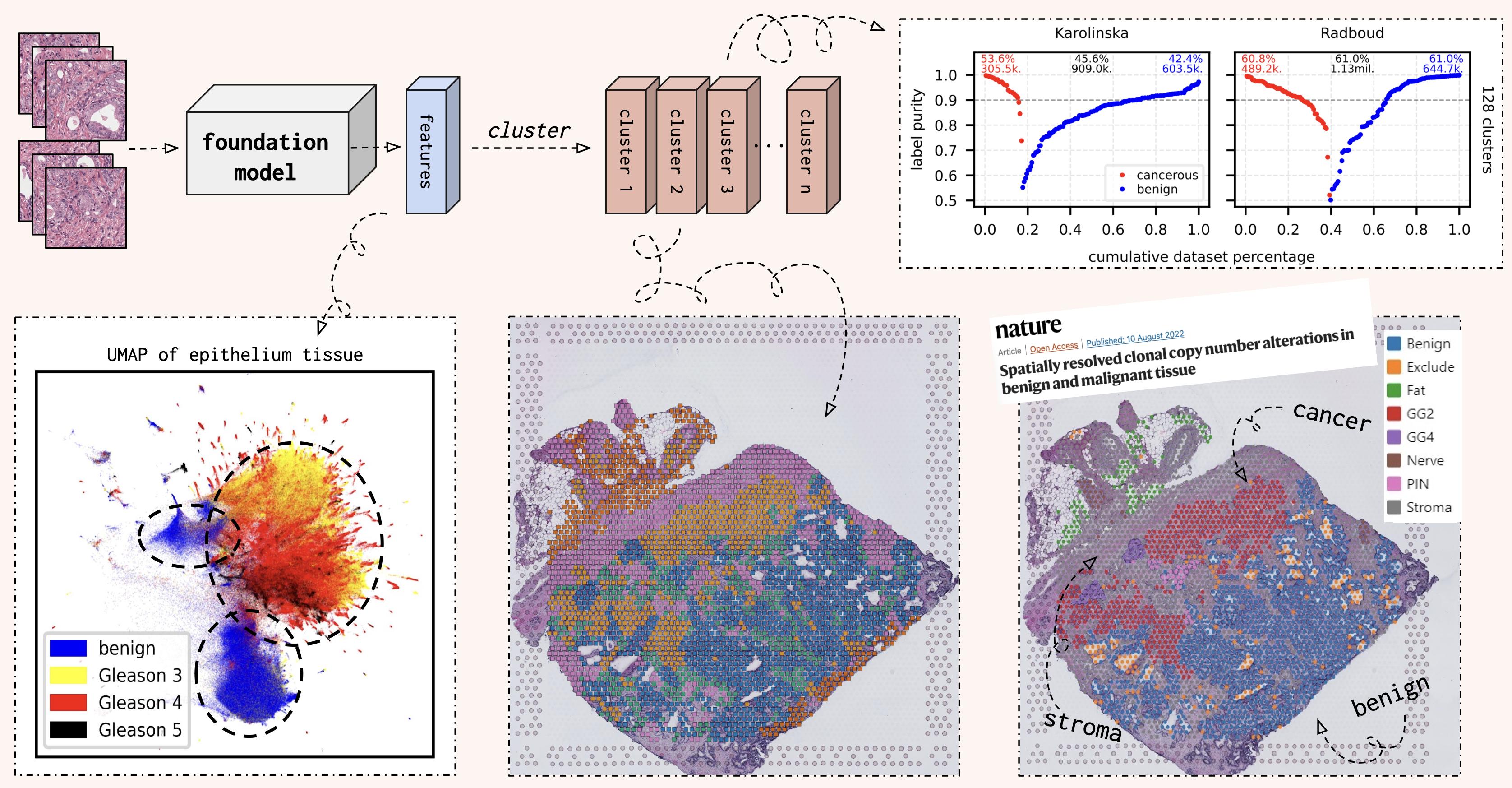
When you feed in tile images (from histological slides), the model will encode these images into a feature vector. When we cluster these features, each cluster will contain tile images with similar histological patterns (cuz that's what the model is good at)! You can do this at a slide level or for a whole dataset to automatically annotate slide images.
In the figure above, you can see UMAP representation of epithelium tissue of the Radboud split of the PANDA dataset. Here we can see that cancerous and benign epithelium form separate clusters, and you can even rediscover the Gleason grading system from the UMAP.
Although UMAPs are pretty, they're kinda shitty for drawing any actual conclusions... Thus, I also clustered all features in the PANDA dataset into 128 clusters. Then we can calculate the propotion of tile images with cancerous or bengin tissue in each cluster. This label purity is visualised on the top right corner, and you can see that ~52% of the 3.8 million tile images are contained in clusters with over 90% label purity!
To learn more, you can check out my slides from the ECDP 2023 seminar. We also have a preprint coming once I finally finish writing it...
Although the first models in this repository (prostate_small, prostate_medium) have
been trained on only prostate tissue, they will very likely be better than any natural
image pre-trained model. Next step of this project/repository is to take the current
models and fine-tune them for other tissues to create other foundation models for new
tissue types (eg. colon_*, breast_* and so on). This fine-tuning step does not
require that much computing power so it's doable even though you don't have access to
thousands of GPUs.
If you're interest in fine-tuning the models for other tissue types, I'm happy to help and share my training scripts/methods! Just send me an email!
pip install histoencoder- Cut histological slide images into small tile images with
HistoPrep.
HistoPrep --input "./slide_images/*.tiff" --output ./tile_images --width 512 --overlap 0.5 --max-background 0.5- Extract features for each tile image.
HistoEncoder extract --input_dir ./tile_images --model-name prostate_small- Cluster extracted features.
HistoEncoder cluster --input_dir ./tile_imagesNow train_tiles contains a directory for each slide with the following contents.
train_tiles
└── slide_image
    ├── clusters.parquet # Clusters for each tile image.
    ├── features.parquet # Extracted features for each tile.
    ├── metadata.parquet # Everything else is generated by HistoPrep.
    ├── properties.json
    ├── thumbnail.jpeg
    ├── thumbnail_tiles.jpeg
    ├── thumbnail_tissue.jpeg
└── tiles [52473 entries exceeds filelimit, not opening dir]If you already have tile images, or want to use your own programs for extracting tile
images, you can use the histoencoder python package! Check out the API
docs for more information.
import histoencoder.functional as F

encoder = F.create_encoder("prostate_small")
for images in your_fancy_dataloader:
   features = F.extract_features(
     encoder, images, num_blocks=2, avg_pool=True
   )
...If you use HistoEncoder models or pipelines in your publication, please cite the
github repository. There's a preprint coming, but I wanted to share the model to the
public as quickly as possible! :)
@misc{histoencoder,
  author = {Pohjonen, Joona},
  title = {HistoEncoder: Foundation models for digital pathology},
  year = {2023},
  publisher = {GitHub},
  journal = {GitHub repository},
  howpublished = {https://github.com/jopo666/HistoEncoder},
}
