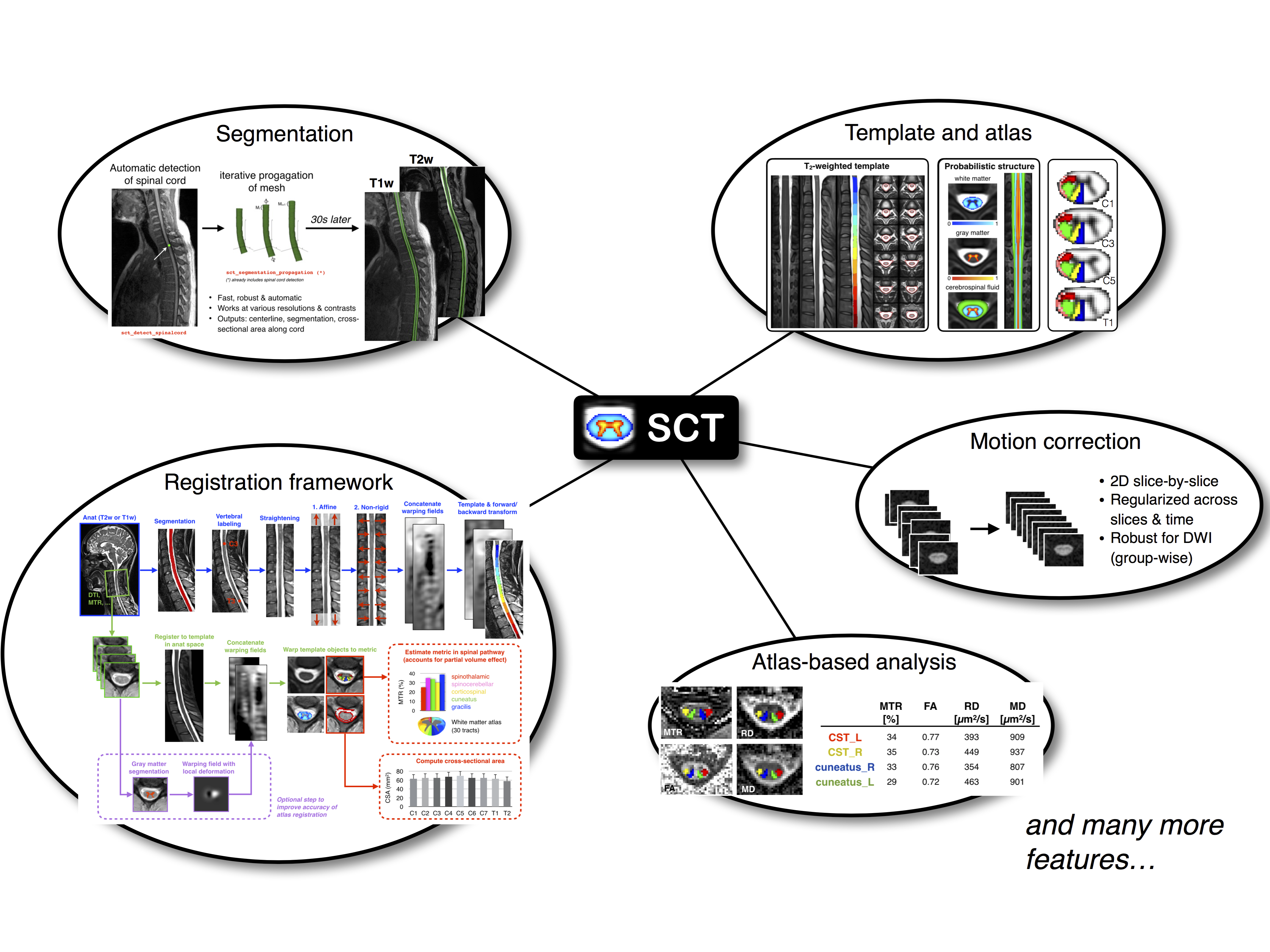
For the past 25 years, the field of neuroimaging has witnessed the development of several software packages for processing multi-parametric magnetic resonance imaging (mpMRI) to study the brain. These software packages are now routinely used by researchers and clinicians, and have contributed to important breakthroughs for the understanding of brain anatomy and function. However, no software package exists to process mpMRI data of the spinal cord. Despite the numerous clinical needs for such advanced mpMRI protocols (multiple sclerosis, spinal cord injury, cervical spondylotic myelopathy, etc.), researchers have been developing specific tools that, while necessary, do not provide an integrative framework that is compatible with most usages and that is capable of reaching the community at large. This hinders cross-validation and the possibility to perform multi-center studies.
Spinal Cord Toolbox (SCT), a comprehensive -source software dedicated to the processing and analysis of spinal cord MRI data. SCT builds on previously-validated methods and includes state-of-the-art MRI templates and atlases of the spinal cord, algorithms to segment and register new data to the templates, and motion correction methods for diffusion and functional time series. SCT is tailored towards standardization and automation of the processing pipeline, versatility, modularity, and it follows guidelines of software development and distribution. Preliminary applications of SCT cover a variety of studies, from cross-sectional area measures in large databases of patients, to the precise quantification of mpMRI metrics in specific spinal pathways. We anticipate that SCT will bring together the spinal cord neuroimaging community by establishing standard templates and analysis procedures.
- Fonov et al. Framework for integrated MRI average of the spinal cord white and gray matter: The MNI-Poly-AMU template. Neuroimage 2014;102P2:817-827.
- Taso et al. Construction of an in vivo human spinal cord atlas based on high-resolution MR images at cervical and thoracic levels: preliminary results. MAGMA, Magn Reson Mater Phy 2014;27(3):257-267
- Lévy et al. White matter atlas of the human spinal cord with estimation of partial volume effect. Neuroimage 2015 (in press); doi: 10.1016/j.neuroimage.2015.06.040
- Cadotte et al. Characterizing the Location of Spinal and Vertebral Levels in the Human Cervical Spinal Cord. AJNR Am J Neuroradiol 2014;36(5):1-8.
- Dupont SM, De Leener B, Taso M, Le Troter A, Stikov N, Callot V, Cohen-Adad J. Fully-integrated framework for the segmentation and registration of the spinal cord white and gray matter. Neuroimage 2016. doi: 10.1016/j.neuroimage.2016.09.026
- De Leener et al. Robust, accurate and fast automatic segmentation of the spinal cord. Neuroimage 2014;98:528-536.
- Ullmann et al. Automatic labeling of vertebral levels using a robust template-based approach. Int J Biomed Imaging 2014;Article ID 719520.
- De Leener et al. Automatic segmentation of the spinal cord and spinal canal coupled with vertebral labeling. IEEE Transactions on Medical Imaging 2015;34(8):1705-1718
- De Leener B, Mangeat G, Dupont S, Martin AR, Callot V, Stikov N, Fehlings MG, Cohen-Adad J. Topologically-preserving straightening of spinal cord MRI. J Magn Reson Imaging 2016. doi: 10.1002/jmri.25622
- De Leener et al. Template-based analysis of multi-parametric MRI data with the Spinal Cord Toolbox. Proc. ISMRM, Toronto, Canada 2015
- Cohen-Adad et al. Slice-by-slice regularized registration for spinal cord MRI: SliceReg. Proc. ISMRM, Toronto, Canada 2015
- Taso et al. A reliable spatially normalized template of the human spinal cord--Applications to automated white matter/gray matter segmentation and tensor-based morphometry (TBM) mapping of gray matter alterations occurring with age. Neuroimage 2015
- Kong et al. Intrinsically organized resting state networks in the human spinal cord. PNAS 2014
- Eippert F. et al. Investigating resting-state functional connectivity in the cervical spinal cord at 3T. Neuroimage 2016
- Weber K.A. et al. Functional Magnetic Resonance Imaging of the Cervical Spinal Cord During Thermal Stimulation Across Consecutive Runs. Neuroimage 2016
- Weber et al. Lateralization of cervical spinal cord activity during an isometric upper extremity motor task with functional magnetic resonance imaging. Neuroimage 2016
- Eippert et al. Denoising spinal cord fMRI data: Approaches to acquisition and analysis. Neuroimage 2016
- Samson et al., ZOOM or non-ZOOM? Assessing Spinal Cord Diffusion Tensor Imaging protocols for multi-centre studies. PLOS One 2016
- Taso et al. Tract-specific and age-related variations of the spinal cord microstructure: a multi-parametric MRI study using diffusion tensor imaging (DTI) and inhomogeneous magnetization transfer (ihMT). NMR Biomed 2016
- Duval et al. In vivo mapping of human spinal cord microstructure at 300mT/m. Neuroimage 2015
- Massire A. et al. High-resolution multi-parametric quantitative magnetic resonance imaging of the human cervical spinal cord at 7T. Neuroimage 2016
- Duval et al. g-Ratio weighted imaging of the human spinal cord in vivo. Neuroimage 2016
- Ljungberg et al. Rapid Myelin Water Imaging in Human Cervical Spinal Cord. Magnetic Resonance in Medicine 2016
- Yiannakas et al. Fully automated segmentation of the cervical cord from T1-weighted MRI using PropSeg: Application to multiple sclerosis. NeuroImage: Clinical 2015
- Martin et al. Next-Generation MRI of the Human Spinal Cord: Quantitative Imaging Biomarkers for Cervical Spondylotic Myelopathy (CSM). Proc. 31th Annual Meeting of The Congress of Neurological Surgeons 2015
- Castellano et al., Quantitative MRI of the spinal cord and brain in adrenomyeloneuropathy: in vivo assessment of structural changes. Brain 2016
- Grabher et al., Voxel-based analysis of grey and white matter degeneration in cervical spondylotic myelopathy. Sci Rep 2016;6:24636.
- Talbott JF, Narvid J, Chazen JL, Chin CT, Shah V. An Imaging Based Approach to Spinal Cord Infection. Semin Ultrasound CT MR. 2016
- McCoy et al. MRI Atlas-Based Measurement of Spinal Cord Injury Predicts Outcome in Acute Flaccid Myelitis. AJNR 2016
- Taso et al. Anteroposterior compression of the spinal cord leading to cervical myelopathy: a finite element analysis. Comput Methods Biomech Biomed Engin 2015
- De Leener et al. Segmentation of the human spinal cord. MAGMA. 2016
- Cohen-Adad et al. Functional Magnetic Resonance Imaging of the Spinal Cord: Current Status and Future Developments. Semin Ultrasound CT MR. 2016
When citing spinalcordtoolbox in academic papers and thesis, please use this BibTeX entry:
@article{DeLeener201724,
title = "SCT: Spinal Cord Toolbox, an open-source software for processing spinal cord \{MRI\} data ",
journal = "NeuroImage ",
volume = "145, Part A",
number = "",
pages = "24 - 43",
year = "2017",
note = "",
issn = "1053-8119",
doi = "https://doi.org/10.1016/j.neuroimage.2016.10.009",
url = "http://www.sciencedirect.com/science/article/pii/S1053811916305560",
author = "Benjamin De Leener and Simon Lévy and Sara M. Dupont and Vladimir S. Fonov and Nikola Stikov and D. Louis Collins and Virginie Callot and Julien Cohen-Adad",
keywords = "Spinal cord",
keywords = "MRI",
keywords = "Software",
keywords = "Template",
keywords = "Atlas",
keywords = "Open-source ",
}
The MIT License (MIT)
Copyright (c) 2014 École Polytechnique, Université de Montréal
Permission is hereby granted, free of charge, to any person obtaining a copy of this software and associated documentation files (the "Software"), to deal in the Software without restriction, including without limitation the rights to use, copy, modify, merge, publish, distribute, sublicense, and/or sell copies of the Software, and to permit persons to whom the Software is furnished to do so, subject to the following conditions:
The above copyright notice and this permission notice shall be included in all copies or substantial portions of the Software.
THE SOFTWARE IS PROVIDED "AS IS", WITHOUT WARRANTY OF ANY KIND, EXPRESS OR IMPLIED, INCLUDING BUT NOT LIMITED TO THE WARRANTIES OF MERCHANTABILITY, FITNESS FOR A PARTICULAR PURPOSE AND NONINFRINGEMENT. IN NO EVENT SHALL THE AUTHORS OR COPYRIGHT HOLDERS BE LIABLE FOR ANY CLAIM, DAMAGES OR OTHER LIABILITY, WHETHER IN AN ACTION OF CONTRACT, TORT OR OTHERWISE, ARISING FROM, OUT OF OR IN CONNECTION WITH THE SOFTWARE OR THE USE OR OTHER DEALINGS IN THE SOFTWARE.


