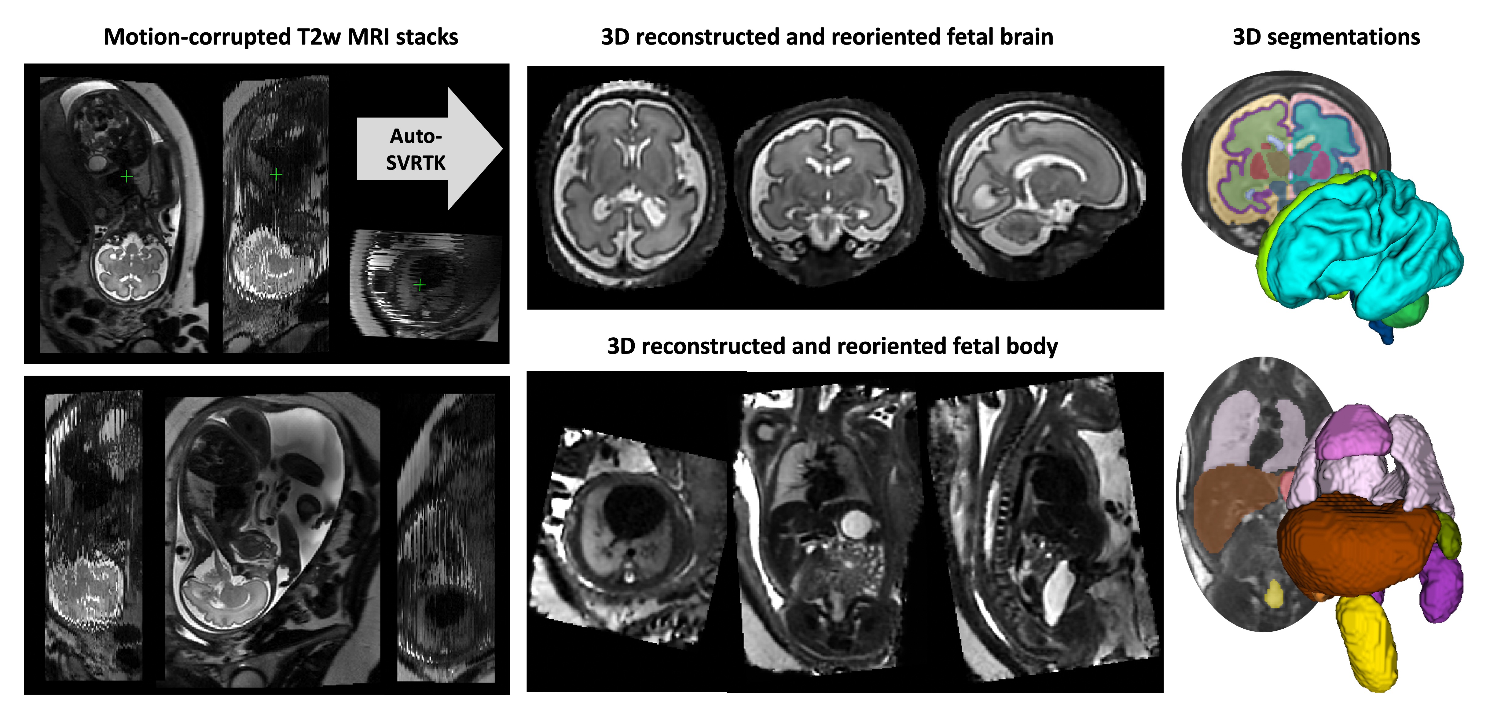
This repository contains the pipelines for MONAI-based automated fetal MRI analysis SVRTK dockers.
- The repository and code for automation of SVR reconstruction and deep learning segmentation were designed and created by Alena Uus, KCL.
The auto pipelines are used in:
-
Integration via AIDE led by Tom Roberts: https://github.com/SVRTK/aide-svrtk
-
Integration via Gadgetron led by Sara Neves Silva: https://github.com/SVRTK/gadgetron-svrtk-integration
Development of SVRTK was supported by projects led by Prof Mary Rutherford, Dr Lisa Story, Dr Maria Deprez, Dr Jana Hutter and Prof Jo Hajnal.
The automated SVRTK docker tag is fetalsvrtk/svrtk:general_auto_amd
AUTOMATED 3D T2w BRAIN / BODY RECONSTRUCTION:
Input data requirements:
- more than 5-6 stacks
- full ROI coverage in all stacks
- 21-40 weeks GA
- no extreme shading artifacts
- singleton pregnancy
- 0.55 / 1.5 / 3T
- 80 – 180ms TE
- sufficient SNR and image quality
Output:
- 0.8mm resolution (or as specified)
- Standard radiological space
docker pull fetalsvrtk/svrtk:general_auto_amd

#auto brain reconstruction
docker run --rm  --mount type=bind,source=LOCATION_ON_YOUR_MACHINE,target=/home/data  fetalsvrtk/svrtk:general_auto_amd sh -c ' bash /home/auto-proc-svrtk/scripts/auto-brain-reconstruction.sh /home/data/folder-with-files /home/data/out-brain-recon-results 1 3.0 0.8 1 ; '

#auto body reconstruction
docker run --rm  --mount type=bind,source=LOCATION_ON_YOUR_MACHINE,target=/home/data  fetalsvrtk/svrtk:general_auto_amd sh -c ' bash /home/auto-proc-svrtk/scripts/auto-body-reconstruction.sh /home/data/folder-with-files /home/data/out-body-recon-results 1 3.0 0.8 1 ; '

#auto thorax reconstruction
docker run --rm  --mount type=bind,source=LOCATION_ON_YOUR_MACHINE,target=/home/data  fetalsvrtk/svrtk:general_auto_amd sh -c ' bash /home/auto-proc-svrtk/scripts/auto-thorax-reconstruction.sh /home/data/folder-with-files /home/data/out-thorax-recon-results 1 3.0 0.8 1 ; '

#auto body reconstruction for CDH
docker run --rm  --mount type=bind,source=LOCATION_ON_YOUR_MACHINE,target=/home/data  fetalsvrtk/svrtk:general_auto_amd sh -c ' bash /home/auto-proc-svrtk/scripts/auto-body-reconstruction-cdh.sh /home/data/folder-with-files /home/data/out-body-recon-results 0 3.0 0.8 1 ; '

#auto head reconstruction
docker run --rm  --mount type=bind,source=LOCATION_ON_YOUR_MACHINE,target=/home/data  fetalsvrtk/svrtk:general_auto_amd sh -c ' bash /home/auto-proc-svrtk/scripts/auto-head-reconstruction.sh /home/data/folder-with-files /home/data/out-head-recon-results 0 3.0 0.8 1 ; '
0.55T low field reconstruction options:
#0.55T auto brain reconstruction
docker run --rm  --mount type=bind,source=LOCATION_ON_YOUR_MACHINE,target=/home/data  fetalsvrtk/svrtk:general_auto_amd sh -c ' bash /home/auto-proc-svrtk/scripts/auto-brain-055t-reconstruction.sh /home/data/folder-with-files /home/data/out-brain-recon-results 1 4.5 1.0 1 ; '

#0.55T auto body reconstruction
docker run --rm  --mount type=bind,source=LOCATION_ON_YOUR_MACHINE,target=/home/data  fetalsvrtk/svrtk:general_auto_amd sh -c ' bash /home/auto-proc-svrtk/scripts/auto-body-055t-reconstruction.sh /home/data/folder-with-files /home/data/out-body-recon-results 1 4.5 1.0 1 ; '
AUTOMATED 3D T2w BRAIN / BODY / ... SEGMENTATION:
Input data requirements:
- sufficient SNR and image quality
- full ROI coverage
- good quality 3D SVR / DSVR reconsruction
- reorientation to the standard radiological atlas space
- 20-38 weeks GA
- no extreme shading artifacts
- no extreme structural anomalies
- 0.55 / 1.5 / 3T
- 80 – 250ms TE
docker pull fetalsvrtk/svrtk:general_auto_amd

#auto brain tissue segmentation
docker run --rm  --mount type=bind,source=LOCATION_ON_YOUR_MACHINE,target=/home/data  fetalsvrtk/svrtk:general_auto_amd sh -c ' bash /home/auto-proc-svrtk/scripts/auto-brain-bounti-segmentation-fetal.sh /home/data/your_folder_with_brain_svr_t2_files  /home/data/output_folder_for_segmentations ; '

#auto brain extraction
docker run --rm  --mount type=bind,source=LOCATION_ON_YOUR_MACHINE,target=/home/data  fetalsvrtk/svrtk:general_auto_amd sh -c ' bash /home/auto-proc-svrtk/scripts/auto-brain-bet-segmentation-fetal.sh /home/data/your_folder_with_brain_svr_t2_files  /home/data/output_folder_for_segmentations ; '

#auto body organ segmentation
docker run --rm  --mount type=bind,source=LOCATION_ON_YOUR_MACHINE,target=/home/data  fetalsvrtk/svrtk:general_auto_amd sh -c ' bash /home/auto-proc-svrtk/scripts/auto-body-organ-segmentation.sh /home/data/your_folder_with_body_dsvr_t2_files  /home/data/output_folder_for_segmentations ; '

#auto lung segmentation (normal and CDH)
docker run --rm  --mount type=bind,source=LOCATION_ON_YOUR_MACHINE,target=/home/data  fetalsvrtk/svrtk:general_auto_amd sh -c ' bash /home/auto-proc-svrtk/scripts/auto-lung-segmentation.sh /home/data/your_folder_with_body_dsvr_t2_files  /home/data/output_folder_for_segmentations ; '

#auto face/head segmentation
docker run --rm  --mount type=bind,source=LOCATION_ON_YOUR_MACHINE,target=/home/data  fetalsvrtk/svrtk:general_auto_amd sh -c ' bash /home/auto-proc-svrtk/scripts/auto-face-segmentation-fetal.sh /home/data/your_folder_with_head_svr_t2_files  /home/data/output_folder_for_segmentations ; '
AUTOMATED REORIENTATION OF 3D T2w BRAIN / BODY / THORAX RECONS TO STANDARD SPACE:
Input data requirements:
- sufficient SNR and image quality
- full ROI coverage
- no extreme shading artifacts
- no extreme structural anomalies
- 0.55 / 1.5 / 3T
- 80 – 250ms TE
docker pull fetalsvrtk/svrtk:general_auto_amd

#auto brain reorientation to the standard space
docker run --rm  --mount type=bind,source=LOCATION_ON_YOUR_MACHINE,target=/home/data  fetalsvrtk/svrtk:general_auto_amd sh -c ' bash /home/auto-proc-svrtk/scripts/auto-brain-reorientation.sh /home/data/your_folder_with_brain_svr_t2_files  /home/data/output_folder_for_reoriented_images 0.5 1 0; '

#auto body reorientation to the standard space
docker run --rm  --mount type=bind,source=LOCATION_ON_YOUR_MACHINE,target=/home/data  fetalsvrtk/svrtk:general_auto_amd sh -c ' bash /home/auto-proc-svrtk/scripts/auto-body-reorientation.sh /home/data/your_folder_with_body_dsvr_t2_files  /home/data/output_folder_for_reoriented_images 0.5 1 0; '

#auto thorax reorientation to the standard space
docker run --rm  --mount type=bind,source=LOCATION_ON_YOUR_MACHINE,target=/home/data  fetalsvrtk/svrtk:general_auto_amd sh -c ' bash /home/auto-proc-svrtk/scripts/auto-thorax-reorientation.sh /home/data/your_folder_with_thorax_dsvr_t2_files  /home/data/output_folder_for_reoriented_images 0.5 1 0; '

#auto body reorientation for CDH cases to the standard space
docker run --rm  --mount type=bind,source=LOCATION_ON_YOUR_MACHINE,target=/home/data  fetalsvrtk/svrtk:general_auto_amd sh -c ' bash /home/auto-proc-svrtk/scripts/auto-body-reorientation-cdh.sh /home/data/your_folder_with_body_cdh_dsvr_t2_files  /home/data/output_folder_for_reoriented_images 0.5 1 0; '
The auto SVRTK code and all scripts are distributed under the terms of the GNU General Public License v3.0. This program is free software: you can redistribute it and/or modify it under the terms of the GNU General Public License as published by the Free Software Foundation version 3 of the License.
This software is distributed in the hope that it will be useful, but WITHOUT ANY WARRANTY; without even the implied warranty of MERCHANTABILITY or FITNESS FOR A PARTICULAR PURPOSE. See the GNU General Public License for more details.
In case you found auto SVRTK useful please give appropriate credit to the software and SVRTK dockers.
Auto brain reconstruction (please include all three citations):
Uus, A. U., Neves Silva, S., Aviles Verdera, J., Payette, K., Hall, M., Colford, K., Luis, A., Sousa, H. S., Ning, Z., Roberts, T., McElroy, S., Deprez, M., Hajnal, J. V., Rutherford, M. A., Story, L., Hutter, J. (2024) Scanner-based real-time 3D brain+body slice-to-volume reconstruction for T2-weighted 0.55T low field fetal MRI. medRxiv 2024.04.22.24306177: https://doi.org/10.1101/2024.04.22.24306177
Uus, A. U., Hall, M., Payette, K., Hajnal, J. V., Deprez, M., Hutter, J., Rutherford, M. A., Story, L. (2023) Combined quantitative T2* map and structural T2- weighted tissue-specific analysis for fetal brain MRI: pilot automated pipeline. PIPPI MICCAI 2023 workshop, LNCS 14246.: https://doi.org/10.1007/978-3-031-45544-5_3
Kuklisova-Murgasova, M., Quaghebeur, G., Rutherford, M. A., Hajnal, J. V., & Schnabel, J. A. (2012). Reconstruction of fetal brain MRI with intensity matching and complete outlier removal. Medical Image Analysis, 16(8), 1550–1564.: https://doi.org/10.1016/j.media.2012.07.004
Auto thorax/body reconstruction (please include both citations):
Uus, A., Grigorescu, I., van Poppel, M., Steinweg, J. K., Roberts, T., Rutherford, M., Hajnal, J., Lloyd, D., Pushparajah, K. & Deprez, M. (2022) Automated 3D reconstruction of the fetal thorax in the standard atlas space from motion-corrupted MRI stacks for 21-36 weeks GA range. Medical Image Analysis, 80 (August 2022).: https://doi.org/10.1016/j.media.2022.102484
Uus, A., Zhang, T., Jackson, L., Roberts, T., Rutherford, M., Hajnal, J.V., Deprez, M. (2020). Deformable Slice-to-Volume Registration for Motion Correction in Fetal Body MRI and Placenta. IEEE Transactions on Medical Imaging, 39(9), 2750-2759: http://dx.doi.org/10.1109/TMI.2020.2974844
Brain tissue segmentation:
Uus, A. U., Kyriakopoulou, V., Makropoulos, A., Fukami-Gartner, A., Cromb, D., Davidson, A., Cordero-Grande, L., Price, A. N., Grigorescu, I., Williams, L. Z. J., Robinson, E. C., Lloyd, D., Pushparajah, K., Story, L., Hutter, J., Counsell, S. J., Edwards, A. D., Rutherford, M. A., Hajnal, J. V., Deprez, M. (2023) BOUNTI: Brain vOlumetry and aUtomated parcellatioN for 3D feTal MRI. eLife 12:RP88818; doi: https://doi.org/10.7554/eLife.88818.1
Body organ segmentation:
Uus, A. U., Hall, M., Grigorescu, I., Avena Zampieri, C., Egloff Collado, A., Payette, K., Matthew, J., Kyriakopoulou, V., Hajnal, J. V., Hutter, J., Rutherford, M. A., Deprez, M., Story, L. (2024) Automated body organ segmentation, volumetry and population-averaged atlas for 3D motion-corrected T2-weighted fetal body MRI. Sci Rep 14, 6637; doi: https://doi.org/10.1038/s41598-024-57087-x
Lung segmentation:
Uus, A., Avena Zampieri, C., Downes, F., Egloff Collado, A., Hall, M., Davidson, J. R., Payette, K., Aviles Verdera, J., Grigorescu, I., Hajnal, J., Deprez, M., Aertsen, M., Hutter, J., Rutherford, M., Deprest, J. & Story, L., (2024). Towards automated multi-regional lung parcellation for 0.55-3T 3D T2w fetal MRI. PIPPI MICCAI Workshop 2024. LNCS vol 14747. https://doi.org/10.1007/978-3-031-73260-7_11
Face segmentation:
Matthew, J., Uus, A., de Souza, L., Wright, R., Fukami-Gartner, A., Priego, G., Saija, C., Deprez, M., Collado, A. E., Hutter, J., Story, L., Malamateniou, C., Rhode, K., Hajnal, J., & Rutherford, M. A. (2024). Craniofacial phenotyping with fetal MRI: a feasibility study of 3D visualisation, segmentation, surface-rendered and physical models. BMC Medical Imaging, 24(1), 52. https://doi.org/10.1186/s12880-024-01230-7
Auto brain reorientation:
Uus, A. U., Hall, M., Payette, K., Hajnal, J. V., Deprez, M., Hutter, J., Rutherford, M. A., Story, L. (2023) Combined quantitative T2* map and structural T2- weighted tissue-specific analysis for fetal brain MRI: pilot automated pipeline. PIPPI MICCAI 2023 workshop, LNCS 14246.: https://doi.org/10.1007/978-3-031-45544-5_3
Auto thorax/body reorientation:
Uus, A., Grigorescu, I., van Poppel, M., Steinweg, J. K., Roberts, T., Rutherford, M., Hajnal, J., Lloyd, D., Pushparajah, K. & Deprez, M. (2022) Automated 3D reconstruction of the fetal thorax in the standard atlas space from motion-corrupted MRI stacks for 21-36 weeks GA range. Medical Image Analysis, 80 (August 2022).: https://doi.org/10.1016/j.media.2022.102484
This software has been developed for research purposes only, and hence should not be used as a diagnostic tool. In no event shall the authors or distributors be liable to any direct, indirect, special, incidental, or consequential damages arising of the use of this software, its documentation, or any derivatives thereof, even if the authors have been advised of the possibility of such damage.

