Problem Statement: Localization of the prostate (healthy or with benign/malignant tumors) in multi-parametric MRI scans (T2W, DWI with high b-value, ADC).
Data (proprietary to Radboud University Medical Center): 1950 prostate mpMRI volumes (Healthy/Benign Cases: 1234; Malignant Cases: 716); equivalent to 23400 2D slices. [1559/391: Train/Val Ratio]
Acknowledgments: The following approach is based on a TensorFlow Estimator/Keras (v1.15) adaptation of keras-retinanet by Fizyr, SEResNet by Pavel Yakubovskiy et al., and an anchor optimization algorithm by Martin Zlocha et al.
Note: The following project is a simplified precursor leading up to the goal of computer-aided clinically significant prostate cancer detection in mpMRI scans, using deep neural network detection models.
Directories
● Preprocess Dataset to Normalized Volumes in Optimized NumPy Format: scripts/preprocess.py
● Generate Data-Directory Feeder List: scripts/feeder_csv.py
● Anchor Optimization: misc/rdc_08.py
● Pre-Calculate Regression Target Deltas *(to determine Mean, STDEV): misc/rdc_07.py
● Train 2D RetinaNet Model: scripts/train_RetinaNet.py
● Deploy Model (Validation): scripts/deploy_model.py
Reference Publications:
● Tsung-Yi Lin et al. (2017), "Focal Loss for Dense Object Detection", IEEE ICCV. DOI:10.1109/ICCV.2017.324
● M. Zlocha et al. (2019), "Improving RetinaNet for CT Lesion Detection with Dense Masks from Weak RECIST Labels", MICCAI. DOI:10.1007/978-3-030-32226-7_45
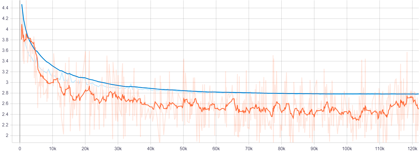
 Figure 1. Training (orange) and validation (blue) loss [focal+L1] curves for the 2D RetinaNet using an exponentially decaying learning rate of 1e-4 with 80% decay every 5 epochs, optimized by SGD with momentum of 0.9.
 Figure 2. Predicted prostate bounding boxes at different scales and orientations [in green] by the 2D RetinaNet, versus the segmentation ground-truth [in blue] (converted to bounding box annotation at train-time) on T2W MRI slices.