When recording electroencephalography (EEG) data, electrodes are usually placed
according to an international standard. The 10-20, and by extension the
10-10 and 10-05 systems are established sets of rules for this case
[1]. Even when
the actual electrode locations have not been empirically measured during the
recording, an approximation of these positions is important for for plotting
topographies or visualizing locations of sensors with the help of analysis
software.
While standard locations are available in many places such as from Robert Oostenveld's blog or directly from electrode cap manufacturers such as Easycap, it is seldom specified how these electrode locations are calculated.
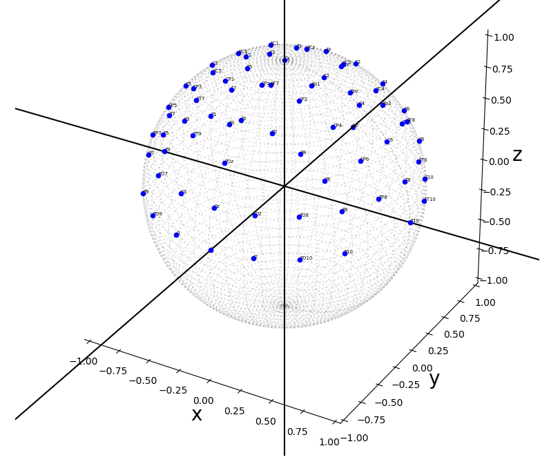
This repository contains code to compute the standard EEG electrode locations
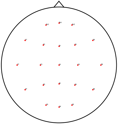
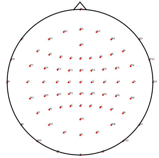
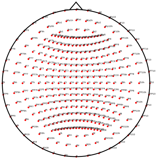
in 3D for the 10-20, 10-10, or even 10-05 system. There are also utility
functions to project the 3D locations to 2D space and plot them.
- The electrode locations are computed on a geometrical sphere centered on the origin and with radius 1 (arbitrary units).
- The function uses an algorithm to compute positions at fractions along
contour lines defined by three points: See the
find_point_at_fractionfunction in theeeg_positions/utils.pyfile.
git clonethe repository (or download as.zipand unpack)cd eeg_positions- Using your python environment of choice, install the package and its
dependencies locally using
pip install -e . - Run the tests using
pytest(you might have topip install pytestfirst) - Calculate and plot electrodes by calling
python eeg_positions/calc_positions.py - Check out
contour_labels.pyfor the order how electrodes are computed - ... and see
utils.pyfor thefind_point_at_fractionfunction that is the core of the computations.
- [1] Oostenveld, R., & Praamstra, P. (2001). The five percent electrode system for high-resolution EEG and ERP measurements. Clinical neurophysiology, 112(4), 713-719. doi: 10.1016/S1388-2457(00)00527-7
Explicit thanks to:
- Robert Oostenveld for writing his blog post on electrodes
- Ed Williams for the helpful correspondence and discussions about "intermediate points on a great circle" (see his aviation formulary)
- "N. Bach" and "Nominal Animal" who helped me to figure out the math for
the
find_point_at_fractionfunction (see this math.stackexchange.com post)
Read the data into MNE-Python
- NOTE: Please download the 3D
10-05data from here and save asstandard_1005.tsv.
import pandas as pd
import numpy as np
import mne

# we saved this file before ...
fname = 'standard_1005.tsv'

# Now read it
df = pd.read_csv(fname, sep='\t')

# Turn data into montage
ch_pos = df.set_index('label').to_dict('index')
for key, val in ch_pos.items():
    ch_pos[key] = np.asarray(list(val.values()))

data = mne.utils.Bunch(
    nasion=ch_pos['Nz'],
    lpa=ch_pos['T9'],
    rpa=ch_pos['T10'],
    ch_pos=ch_pos,
    coord_frame='unknown',
    hsp=None, hpi=None,
)

montage = mne.channels.make_dig_montage(**data)

# plot it
montage.plot()reproduce by running python eeg_positions/calc_positions.py
reproduce by running python eeg_positions/calc_positions.py
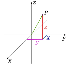
- Imagine the x-axis pointing roughly towards the viewer with increasing values
- The y-axis is orthogonal to the x-axis, pointing to the right of the viewer with increasing values
- The z-axis is orthogonal to the xy-plane and pointing vertically up with increasing values
For simplicity, we assume a spherical head shape of a human. Roughly speaking, the x-axis goes from the left ear through the right ear, the y-axis goes orthogonally to that from the inion through the nasion, and the z-axis goes orthogonally to that plane through the vertex of the scalp.
We use the following anatomical landmarks to define the boundaries of the sphere:
- The left preauricular point =
(-1, 0, 0)... coincides with T9 - The right preauricular point =
(1, 0, 0)... coincides with T10 - The nasion =
(0, 1, 0)... coincides with Nz - The inion =
(0, -1, 0)... coincides with Iz - The vertex =
(0, 0, 1)... coincides with Cz
Hence, the equator of the sphere goes through T9, T10, Nz and Iz.
Note that these are ASSUMPTIONS. It would be equally valid to assume the equator going through T7, T8, Fpz, and Oz.
You can also just download the pre-computed electrode positions in tab-separated data format.
