The need for image analysis is ever growing in many fields and cancer is not an exception. With the advent of new imaging techniques such as intravital, confocal and multiphoton microscopy, just to mention a few, researchers can visualise physiological and pharmacological processes together with the traditional anatomical images that have been improving resolution. Yet, once the imaging of subjects has been achieved, sometimes there is a lack of resources to properly analyse and process the data and the wealth of the information contained in images and videos remains to be extracted in the near future. The development of methods of analysis that extract meaningful and quantitative information from these images is an important part in the search of understanding biological processes
CAIMAN originated as an Image Analysis internet-based project that combined the strength
of open-source web-based scripting languages, the powerful
high-level technical computing language MATLAB,
and the vast literature on image analysis and computer vision to
provide a user-friendly web-page where any person can upload
cancer-related images and execute analysis algorithms and obtain
quantitative measurements related to their images.
With time, it become evident that for some people it may be better to test and adapt the algorithms as Matlab Scripts to be run locally instead of cloud-based. The algorithms available at CAIMAN can now be accessed in GitHub, however, the cloud-based system is still running at:
For the Matlab routines follow these links:
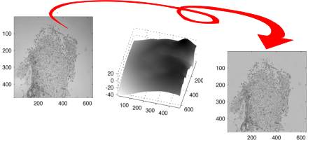
- shading correction based on a signal envelope estimation retrospective algorithm,
https://github.com/reyesaldasoro/shading-correction
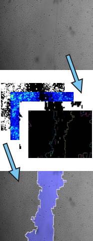
- measuring cellular migration for scratch wound assays
https://github.com/reyesaldasoro/Cell-Migration
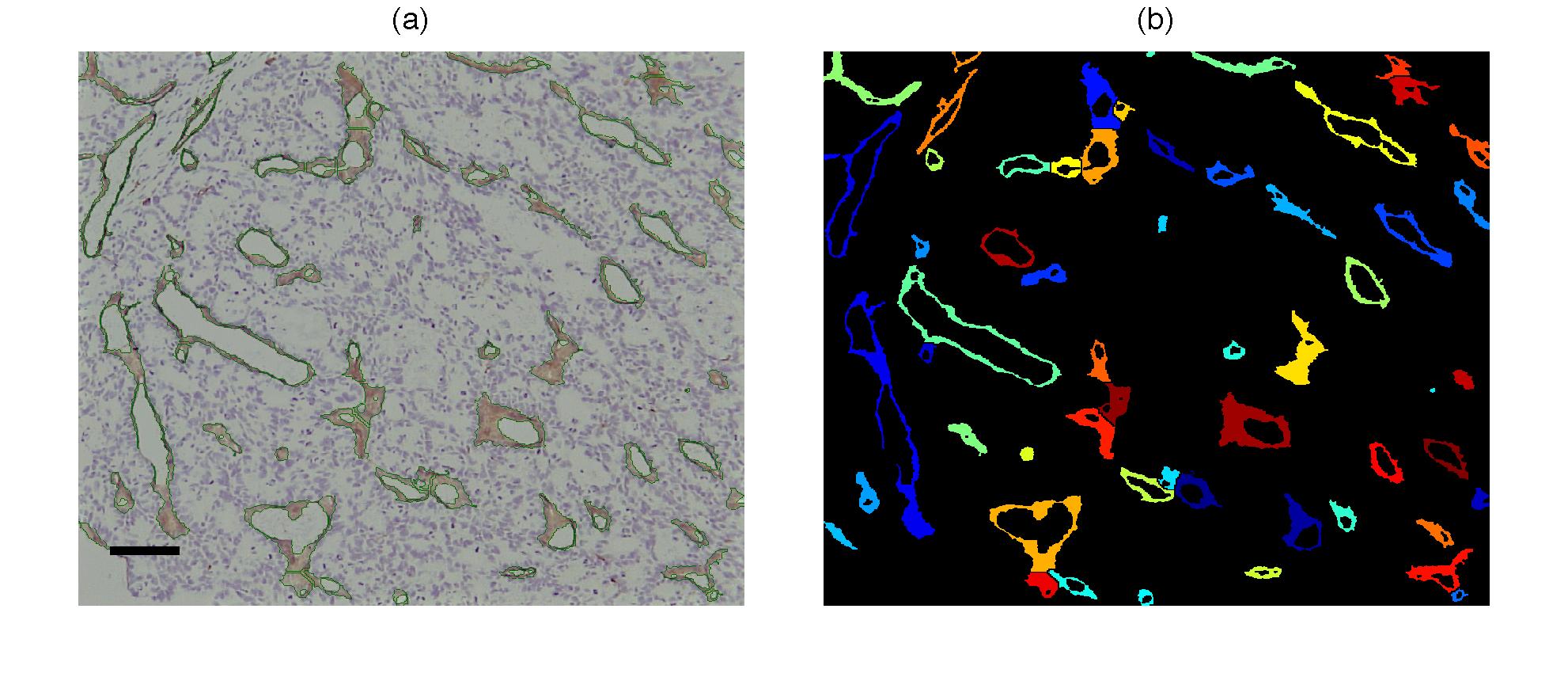
- Microvessel Segmentation from tissue stained with immunohistochemistry (CD-31, blue-brown)
https://github.com/reyesaldasoro/Microvessel-Segmentation
- Tracing of vessels for in-vivo intravital microscopy
https://github.com/reyesaldasoro/Scale-Space-Vessel-Tracing
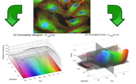
- Chromatic Analysis originally for immunohistochemistry and intravital microscopy, but can be used for anything


