Machine learning, perception and discovery Lab
Mathematician and Master in Applied Mathematics by the National University of Colombia. Ph.D. student in Systems and Computing Engineering. More than five years of experience in teaching in higher education. Experience in the design and implementation of algorithms and software. More tan one year of research experience, member of the MindLab (Machine learning, perception and discovery Lab) research group. Active research in Machine Learning.
Computational Learning Model for the Eye Fundus Analysis to Support Medical Diagnosis
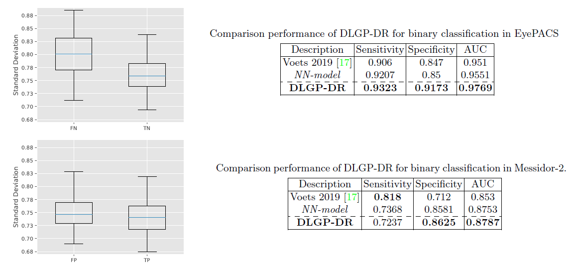
Toledo-Cortés, S., De La Pava, M., Perdomo, O., and González, F.A. (2020) Hybrid Deep Learning Gaussian Process for Diabetic Retinopathy Diagnosis and Uncertainty Quantification, arXiv preprint.
[ENG]
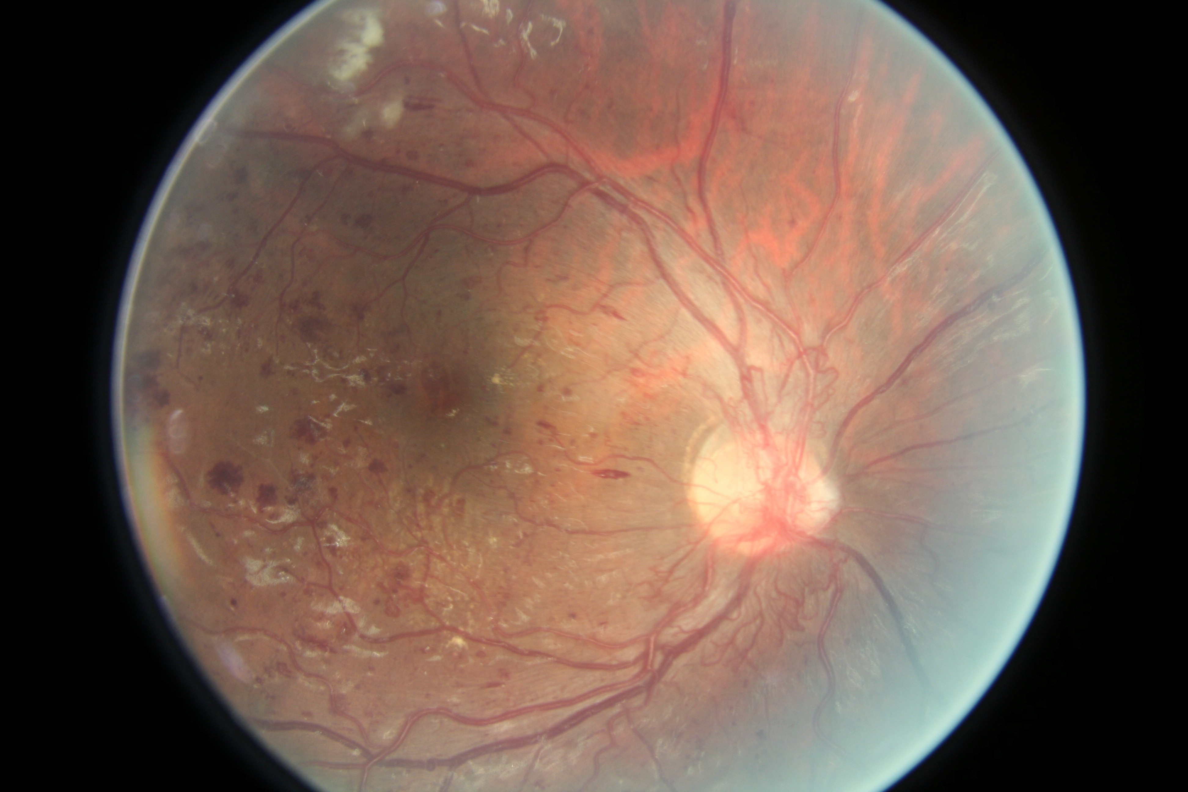
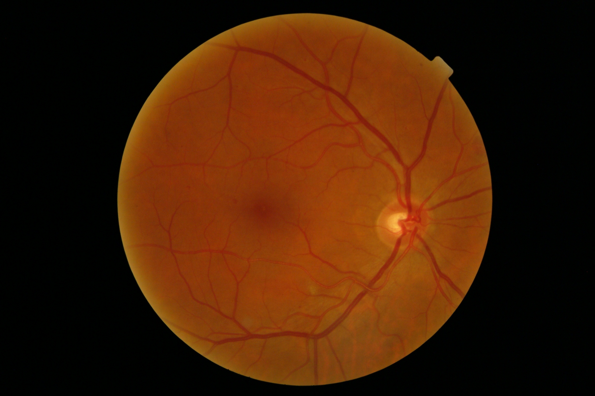
- Color photo of the posterior pole of the right eye, slight opacity of the media, a disc with well-defined edges, with slight pallor. The presence of neovascularization is observed at the level of the optic nerve that exists extending to the upper and lower arches. Multiple points and flame hemorrhages are observed throughout the posterior pole, associated with the presence of exudation in the central macular region. Multiple cottony white spots are also seen on the super-temporal aspect. Diagnosis: Proliferative diabetic retionpathy and moderate diabetic macular edema.
[SPA]
- Foto color de polo posterior del ojo derecho, opacidad leve de medios, disco de bordes bien definidos, con leve palidez. Se observa la presencia de neo-vascularización a nivel del nervio óptico que existe que se extiende hacia arcadas superiores e inferiores. Se observa en todo el polo posterior múltiples hemorragias en punto y en llama asociado a presencia de exudación en la región macular central. También se observan múltiples manchas blancas algodonosas sobre el aspecto superior-temporal. Diagnóstico: Retinopatía diabética proliferativa y edema macular diabético moderado.
-
Presence of: Aneurysms, Hemorrhages and Cotton-wool spots.
-
No presence of: Venous beading, Neovascularization and Exudates.
Diagnosis: Referable patient
To download the dataset please send an email address to stoledoc@unal.edu.co. I will send you the information on how to obtain the dataset once it's ready to be downloaded.


